Figure 6.
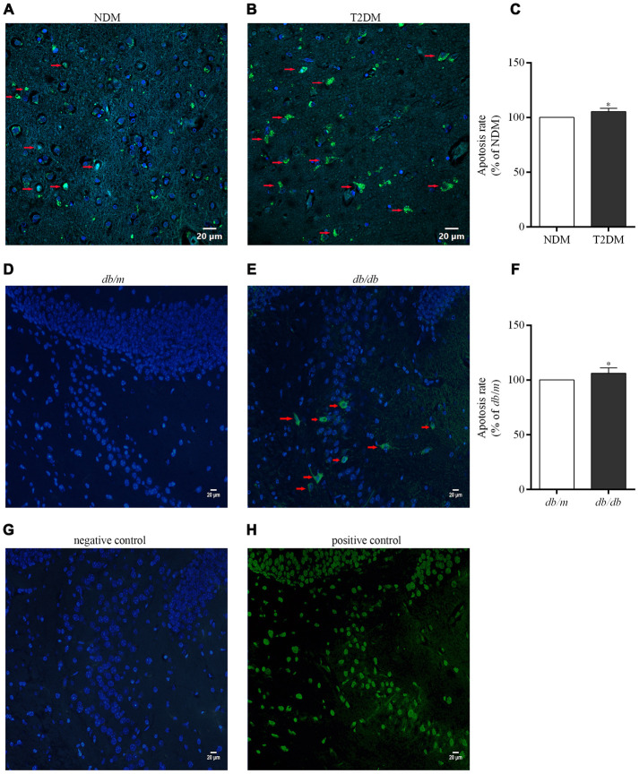
Apoptotic cells in the brains of the patients with T2DM and db/db mice as well as controls in situ detected with the TUNEL assay. Photographs were taken by using a laser confocal microscope (scale bars=20 μm). The co-localization of nuclei (DAPI, blue) and TUNEL-positive cells (green) indicated by red arrow are shown in the merged images. (A) NDM; (B) T2DM; (C) apoptosis rate of neurons in the human brains; (D) db/m mice; (E) db/db mice; (F) apoptosis rate of neurons in the mouse brains; (G) negative control for the method; (H) positive control for the method. The values presented as percentage of the control by relative quantification are mean ± SEM (n=5 for each group). *p<0.05 as compared to NDM (C) or db/m mice (F) as determined by the two-tailed unpaired Student’s t test.

