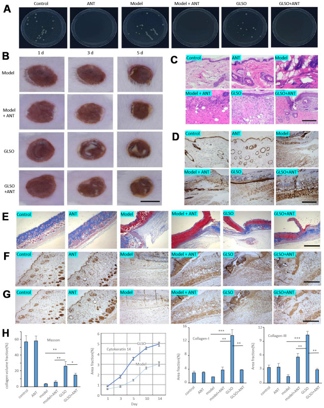
Figure 6.
GLSO accelerates the skin wound healing more rapidly than the group with treatment of antibiotics. (A) The culture of skin microbiota on each group after 7 days with the treatment of antibiotics. (B) Gross examination of the wound area on each group. Scale bar =6 mm. (C) H&E staining of the wound tissue sections. (D) Immunohistochemical analysis of cytokeratin 14 under the microscope 100X, scale bar=400 μm. (E) Masson’s Trichrome staining, scale bar=1 mm. Immunohistochemical analysis of Collagen I (F) and Collagen III (G) at the murine skin wound on each group under the microscope 100X, scale bar=0.4 mm. (H) Quantitative analysis of Masson’s Trichrome staining, and immunostaining of cytokeratin 14 (quantitation results of Fig 1E), Collagen I, and Collagen III (n=3 each group). The data are indicated as the mean±SD. *P<0.05,**P <0.01, ***P <0.001(*P: other groups vs GLSO).

