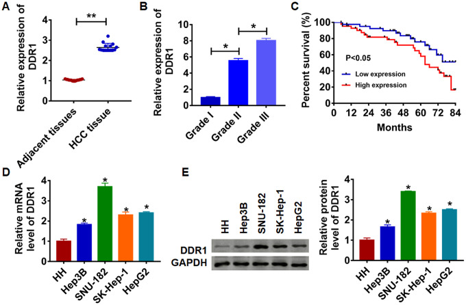
Figure 1.
Expression of DDR1 in HCC tissue and cells. (A) The expression of DDR1 in clinical HCC tissues (n = 15) and adjacent normal tissues (n = 15) determined by qRT-PCR (**p<0.01). (B) Another HCC tissues from patients with tumor grade I (n = 8), grade II (n = 8) and grade III (n = 8) were collected, and the expression of DDR1 in was measured by qRT-PCR (*p<0.05). (C) The overall survival of HCC patients with low or high expression of DDR1 in HCC tissues were assessed by Kaplan-Meier survival analysis (n = 15, *p<0.05). qRT-PCR assay (D) and western blot (E) analyzed the expression of DDR1 in human hepatocyte (HH) and HCC cell lines Hep3B, HepG2, SK-Hep-1 and SNU-182 (n = 6, *p<0.05 vs HH).

