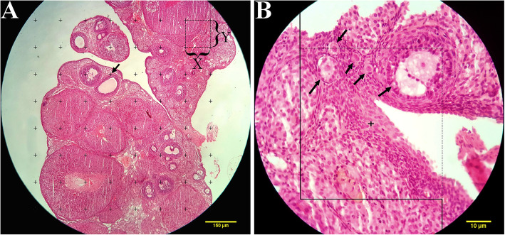
Fig. 1.
a Point-counting method to estimate the volume density of the cortex, medulla, cystic formation, and corpus luteum. The arrow indicates the right upper quadrant of each cross that was considered to be a point. b The optical disector method. An unbiased counting frame was laid on the images. The follicle oocytes were counted if they placed completely or partly inside the counting frame, touched the upper and right lines (acceptance line), or did not touch the left and lower borders (rejection lines) (arrows). Upper and lower guard zones were defined, and each of them was set to be 5 μm using a Heidenhain microcator (MT12, Germany)

