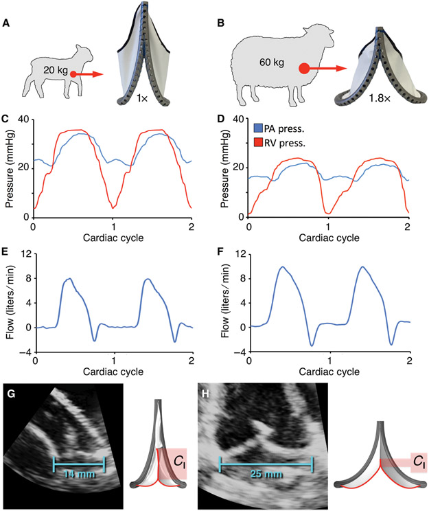
Fig. 4. Acute in vivo validation of primary biomimetic valve function in juvenile and adult sheep.
(A and B) Fabricated prototypes implanted at two polar expansion states (1X and 1.8X) in the native pulmonary valve position of juvenile (N = 4) and adult (N = 4) sheep. Representative plots showing right ventricular and pulmonary artery pressures for the (C) 1X and (D) 1.8X valve geometries. Corresponding pulmonary artery flow for the (E) 1X and (F) 1.8X valve geometries. Representative echocardiographic images of implanted valves, demonstrating change in coaptation length (Cl) between the (G) baseline (1X, 14 mm ID) and (H) fully expanded (1.8X, 25 mm ID) geometries. ID = internal diameter.

