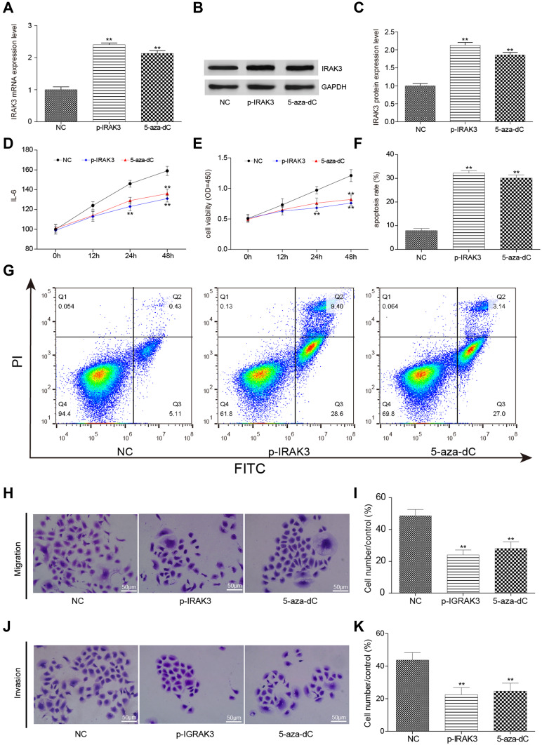
Figure 7.
Effect on glioma cells after the overexpression and demethylation of the IRAK3. (A) The expression level of the IRAK3 was significantly higher in the glioma U251 cells of the pcDNA3.1-IRAK3 and 5-aza-dC groups than in the control group. (B and C) The protein expression level of the IRAK3 significantly increased in the pcDNA3.1-IRAK3 and 5-aza-dC groups compared with the control group. (D) The expression level of the inflammatory factor IL-6 significantly decreased in the pcDNA3.1-IRAK3 and 5-aza-dC groups compared with the control group. (E) The activity significantly decreased in the pcDNA3.1-IRAK3 and 5-aza-dC groups compared with the control group. (F and G) The apoptosis rate significantly increased in the pcDNA3.1-IRAK3 and 5-aza-dC groups compared with the control group. (H and I) The migration capability was significantly lower in the pcDNA3.1-IRAK3 and 5-aza-dC groups compared with the control group. (J and K) The invasiveness capability was significantly lower in the pcDNA3.1-IRAK3 and 5-aza-dC groups than in the the control group. All the mentioned differences were significant (**P < 0.01). The IRAK3 had a suppressive effect on glioma cells in vitro.

