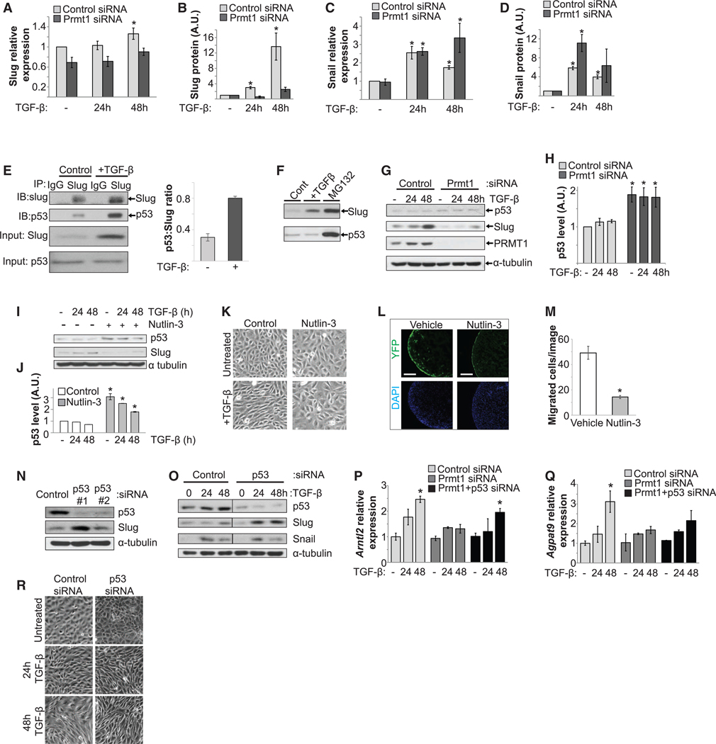
Figure 5. Silencing PRMT1 Increases the p53 Level to Reduce Slug Induction.

(A–D) Slug induction during epicardial EMT occurred mainly at the protein level, in contrast to Snail. TGF-β induced a modest increase in mRNA levels of Slug (A) but a dramatic increase in protein expression (B), in contrast to a dramatic increase in both mRNA (C) and protein levels (D) for Snail. mRNA was measured by qRT-PCR, and protein expression was quantified by immunoblot from multiple assays. *p < 0.05 versus untreated.
(E) Slug interacted with p53 in MEC1 cells. Co-immunoprecipitation (coIP) using anti-Slug antibody or immunoglobulin G (IgG) control was blotted for Slug andp53, and quantification for the p53:slug ratio. IB, immunoblot. IP, immunoprecipitation. The input images were the same as the first two columns of (F) from the same assay.
(F) Slug and p53 were degraded by the proteasome. The protein levels of Slug and p53 were increased by MG132, which blocks proteasomal degradation.
(G and H) Silencing Prmt1 increased p53 protein levels. p53 is blotted in (G), and the blot is quantified in (H). *p < 0.05 versus control siRNA.
(I and J) Enhancing p53 levels with Nutlin-3 reduced TGF-β-induced Slug in MEC1 cells, as assessed by immunoblotting in (I) and quantified in (J). *p < 0.05 versus control.
(K) Nutlin-3 prevented EMT morphological change, as assessed by phase contrast microscopy.
(L and M) Nutlin-3 decreased epicardial invasion. Hearts dissected from E12.5 Wt1-CreERT;ROSAYFP/+ embryos were incubated with vehicle or Nutlin-3 in ex vivo culture. YFP-labeled epicardial-derived cells in the myocardium are visualized in (L) and quantified in (M). The left ventricle is shown. *p < 0.05 versus vehicle.
(N) Depletion of p53 increased Slug levels. MEC1 cells transfected with control or two independent p53 siRNAs were assessed.
(O) Depletion of p53 with siRNA 1 enhanced Slug induction, but not Snail, during epicardial EMT.
(P and Q) PRMT1-p53 pathway regulated Slug target gene induction. mRNA levels of Slug target genes Arntl2 (P) and Agpat9 (Q) were analyzed by qRT-PCR. *p < 0.05 versus control siRNA.
(R) Depletion of p53 accelerated EMT morphological change, as assessed by phase contrast microscopy.
