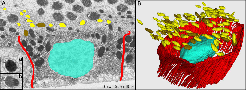
Figure 2.
Melanosomes and lipofuscin granules in RPE apical processes. (A) Electron-microscopy cross-section with inset a showing a representative melanosome and inset b a lipofuscin granule. Cell borders of one RPE cell are highlighted in red. (B) Manually reconstructed 3D view of one RPE cell with associated complete reconstructions of melanosome and lipofuscin granules located in the apical processes (not shown). Colors: red, RPE cell borders; turquoise, cell nucleus; yellow, melanosomes; brown, lipofuscin.

