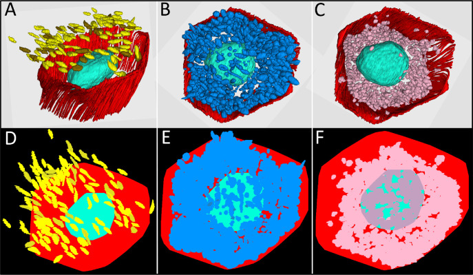
Figure 6.
RPE cell coverage area of reflective granules and mitochondria. The top row shows 3D reconstructions of melanosome (yellow) and lipofuscin (brown) granules in the apical processes of RPE cells (A), combined population of lipofuscin, melanolipofuscin, and melanosome granules (blue) in the RPE cell body (B), and mitochondria (pink) in the RPE cell body (C). RPE cell borders are indicated in red and cell nucleus in turquoise. The bottom row (D–F) indicates the modeled coverage area of the respective granules and mitochondria (see color code for A–C) visualized from an apical view. (D) Coverage area of melanosomes and lipofuscin granules in RPE apical processes with some granules outside the cell borders due to bending of apical processes occurring during processing of tissue. (E) Reflective granules (lipofuscin, melanolipofuscin, melanosome) and (F) mitochondria in the cell body with dark pink areas indicating mitochondria located on the basal side of the nucleus.

