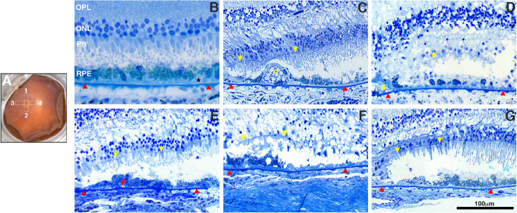
Figure 2.
Histologic morphology of RPE and retina in GA borders of donors with AMD. Fundus image of a control eye (A, OD) with a schematic drawing of the GA areas harvested and processed for further analysis. These include the superior (region 1), nasal (region 4), temporal (region 3), and inferior (region 2). Toluidine blue–stained plastic sections (1 µm) of retinas from all AMD donors and an age-similar control. Morphologic study of the control retina (B) displayed typical retinal lamina, slightly “nonuniform” RPE structure, and pigmentation with small patches of early basal laminar deposit (BLamD, *) over the Bruch's membrane (red arrowheads). Histologic study in the edge of the GA atrophy of AMD donors displayed degenerated photoreceptors and RPE. Observed RPE degeneration morphology included “very nonuniform” RPE (C), “dissociated” RPE (D), “shedding” RPE (E); “bilaminar” RPE (F), and “sloughed” RPE (G). Yellow arrows show ELM (external limiting membrane) descending toward the Bruch's membrane to define the GA border; red arrows show shed RPE contents. D, drusen. Scale bar: 100 µm.

