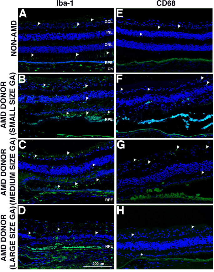
Figure 5.

Immunocytochemistry of retinal sections in the GA borders of donors with AMD stained with inflammatory cell marker antibodies. Immunofluorescence of control and AMD retinal sections labeled with antibodies to Iba-1 (A, B, C, D) and CD68 (E, F, G, H). Non-AMD donor displayed very few Iba-1+ cells in their retinas, with cells mostly localized in the inner retina, but there were a few also localized to the photoreceptor outer segments (A, white arrowheads). The AMD donors with small and medium size GA atrophy (B, C) displayed infiltration of Iba-1+ cells in the outer retina; these cells displayed an amoeboid phenotype. However, fewer Iba-1+ cells were present in the retinas of AMD donors with big size GA atrophy (D). Non-AMD donor (E) displayed very few CD68+ cells in their retinas. The AMD donors with small and medium size GA atrophy displayed increased presence of CD68+ cells in the inner retina (F, G, white arrowheads). However, CD68+ cells were mostly absent from AMD donors with big size GA atrophy (D). For all images, the atrophy was located at the left edge of the picture. Scale bar: 200 µm.
