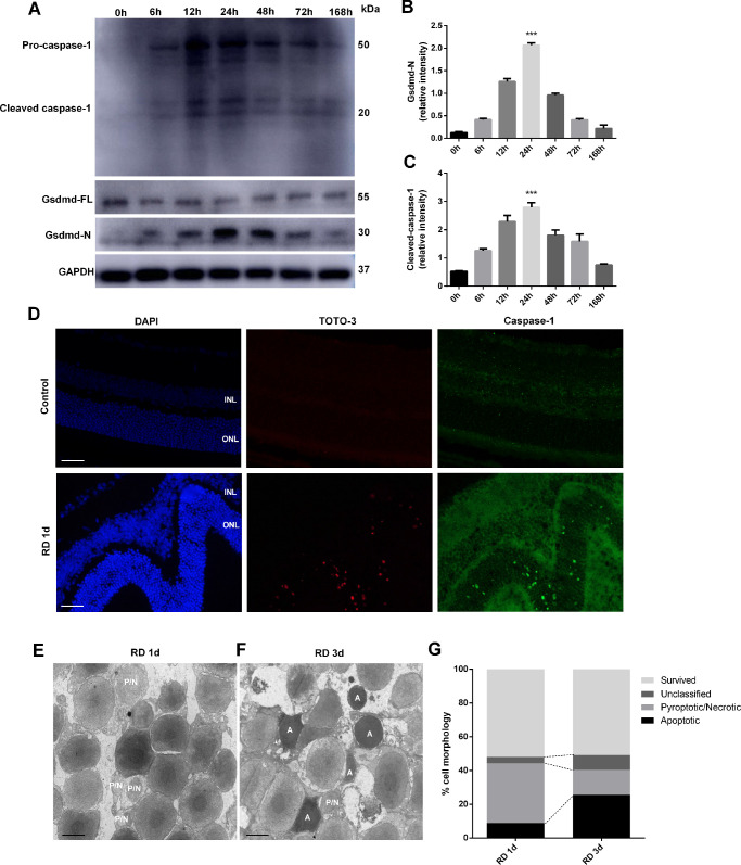
Figure 1.
GSDMD and caspase-1 expression, TEM of photoreceptors 1 day and 3 days after RD, and TOTO-3 and caspase-1 staining 1 day after RD. (A) Time course of GSDMD-N, GSDMD-FL, and cleaved caspase-1 levels in rat retina, detected by western blotting at 0 hour (control eyes, n = 6) and from 6 to 168 hours after RD surgery (n = 6 per time point). (B, C) Densitometry analysis of the western blotting data normalized to the intensity of GAPDH (n = 6). Statistical analyses were performed via ANOVA with Bonferroni correction for the three independent experiments. Data are presented as the mean ± SD. (D) Representative TOTO-3 and caspase-1 staining of photoreceptors 1 day after RD (n = 6). Scale bar: 25 µm. (E, F) Representative TEM photographs of photoreceptors 1 day and 3 days after RD (n = 6). Scale bar: 2 µm. (G) Quantification of different cell death forms 1 day and 3 days after RD. ***P < 0.001. INL, inner nuclear layer; P/N, pyroptotic/necrotic; A, apoptotic.

