Figure 5.

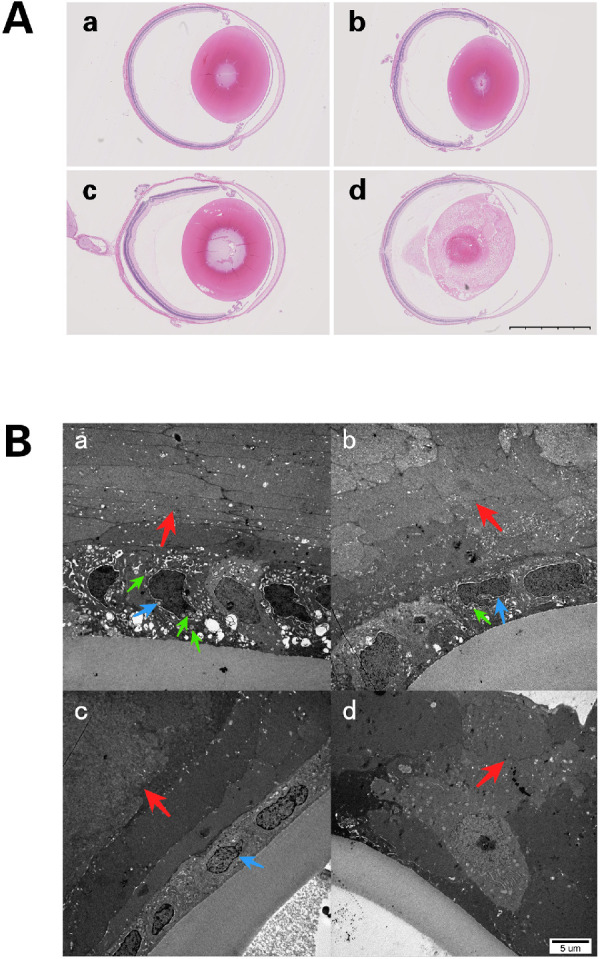
(A) Lens sections with H&E staining at P21. (a) Lenses from the control group were normal in appearance, (b) lenses from the 50-mg/kg group were nearly normal in appearance. (c) Liquefied fibers were found in the posterior pole in rats from the 60-mg/kg group. (d) Mature cataract formation in lenses from the 70-mg/kg group. Scale bar = 2.5 mm. (B) Transmission electron microscopy examination at P14. (a) Samples from the control group show normal LECs with normal nuclei (blue arrow), abundant mitochondria (green arrow) in the cytoplasm, and organized fibers (red arrow). (b) In the 50-mg/kg group, LECs have slightly shrunken cytoplasm and nuclei (blue arrow) with limited numbers of mitochondria (green arrow) and fibers showing slight swelling and lack of organization (red arrow). (c) In the 60-mg/kg group, LECs show deeply shrunken cytoplasm with condensation and dysmorphism of the nuclear chromatin (blue arrow). The subepithelial fibers of these LECs were swollen and disorganized (red arrow). The number of mitochondria in these LECs decreased markedly. (d) In the 70-mg/kg group, the LECs have disappeared, and the fibers are markedly swollen and disorganized (red arrow).
