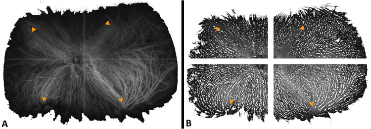
Figure 1.
Representative mid-phase ultra-widefield indocyanine green angiography images (Optos California; Optos, Inc., Dunfermline, UK) after removal of imaging artifacts from a left eye diagnosed with chronic central serous chorioretinopathy. (A) Using multilevel image thresholding with Otsu’s method47 and local adaptive threshold calculations based on pixel values and (B) after segmentation to quadrants based on the most central extension of the choroidal vessel draining into respective vortex veins (yellow arrowheads).

