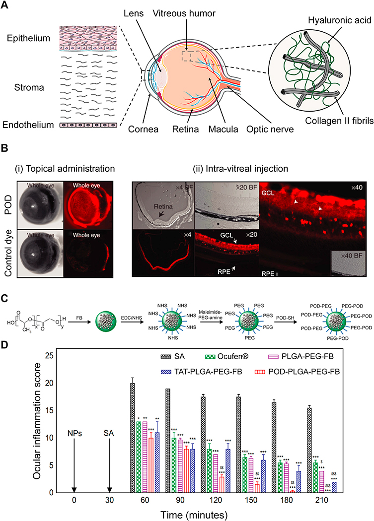
Fig. 5.
A. Structure of ocular tissue in the anterior (cornea and the lens) and posterior (vitreous humor, retina and macula) regions. Barriers to local drug delivery include negatively charged corneal epithelium layer and vitreous humor comprising of collagen II and hyaluronic acid that prevent diffusion of drugs and their carriers to the posterior regions. B. Transport of POD in ocular tissues upon (i) topical administration and (ii) intra-vitreal injection. (i) POD was uptaken by external ocular tissues within 45 minutes upon topical administration in mice eyes while the control free dye only weakly stained the eye. (ii) Intra-vitreal injection of POD transduced 85 % of neural retina within 2 h. RPE and GCL represent retinal pigment epithelium and ganglion cell layer, respectively. Adapted from ref [137]. Printed with permission from Elsevier. C. Synthesis of POD-PLGA-PEG-FB nanoparticles. D. Effectiveness of POD-PLGA-PEG-FB in prevention of sodium arachidonate (SA) induced ocular inflammation in rabbit eyes (*p < 0.05, **p < 0.01, and ***p < 0.001 vs inflammation induced by SA.$p< 0.05, $ $p< 0.01, and $ $ $p < 0.001 vs anti-inflammatory effect of Ocufen®). Adapted from ref [140]. Printed with permission under Creative Commons Attribution License.

