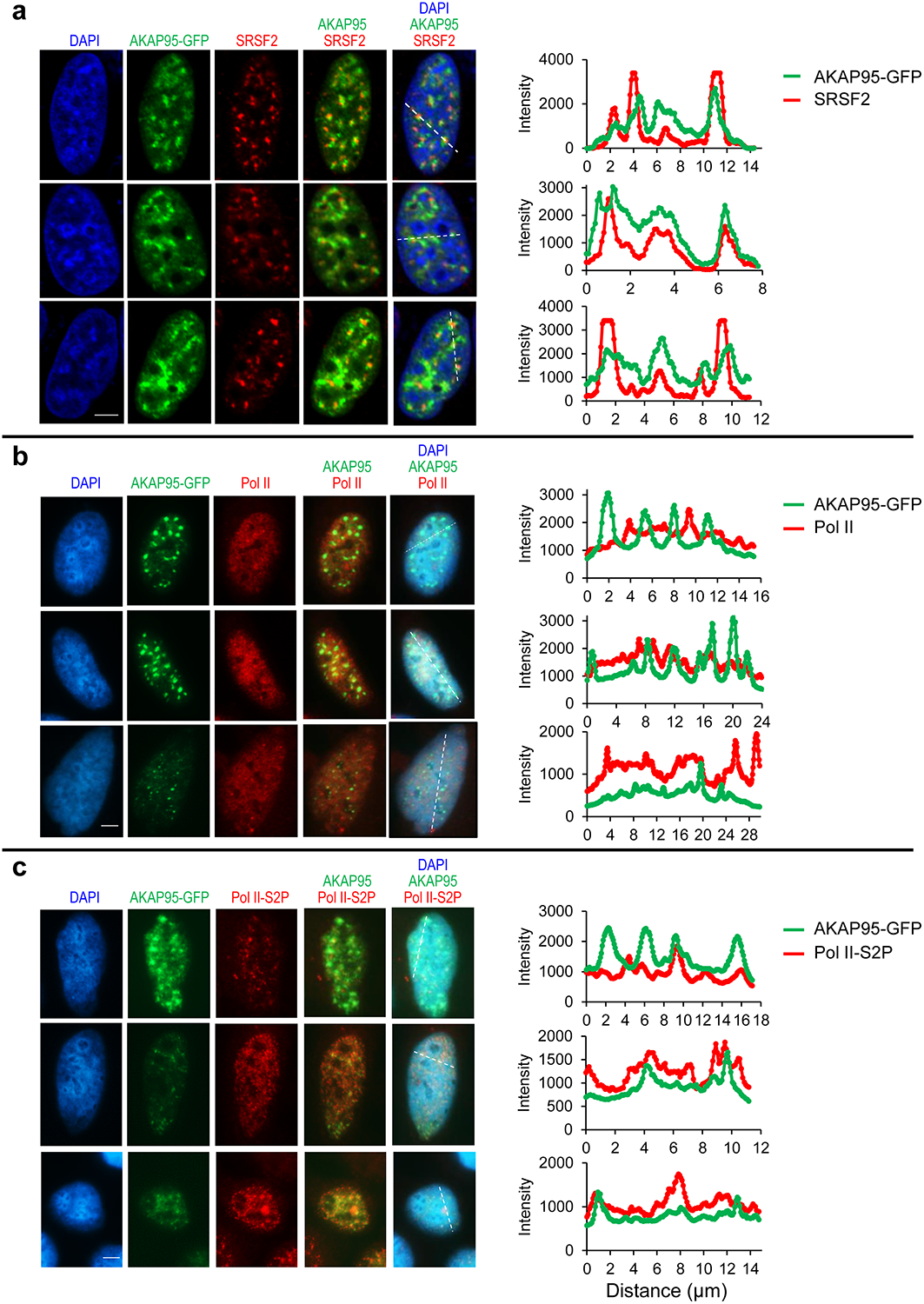
Extended Data Fig. 5. AKAP95 partially localizes in nuclear speckles and with actively transcribing Pol II.

Fluorescence microscopy images of HeLa cells transiently expressing AKAP95-GFP. Nucleus DNA was stained by DAPI, and specific proteins were stained with antibodies for SRSF2 (a), Pol II (b), and Pol II-S2P (c). The assays were repeated 10 times for each staining and show similar trend. Right, quantification of the signal intensity of indicated molecules across the dotted lines shown in the images. Quantification by Image J. Scale bar, 5 μm for all. Statistical source data are provided as in source data extended data fig 5.
