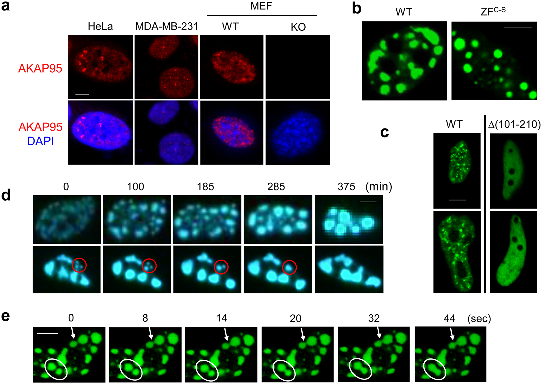
Fig. 5. AKAP95 forms dynamic foci in cell nucleus.

a, Immunostaining of endogenous AKAP95 (red) and DNA (DAPI, blue) in indicated cancer cell lines and primary MEFs from WT and Akap95 KO embryos.
b, Confocal microscopy images of AKAP95 WT or ZFC-S fused to GFP in nuclei following transfection into HeLa cells.
c, Fluorescence microscopy images of HeLa cells transiently expressing AKAP95 WT or Δ(101–210 fused to GFP.
d, HeLa cells were transfected with AKAP95-GFP, and two nuclei were imaged at different time points. Time 0 was 24 hr after transfection. Note the growth and merge of the foci, especially those in the red circle.
e, Rapid fusion of AKAP95 (ZFC-S)-GFP foci in a HeLa cell nucleus. The white oval and arrow show two different fusion events. These images are from Video 2.
All experiments were Repeated 4 times. Scale bar, 5 μm for all.
