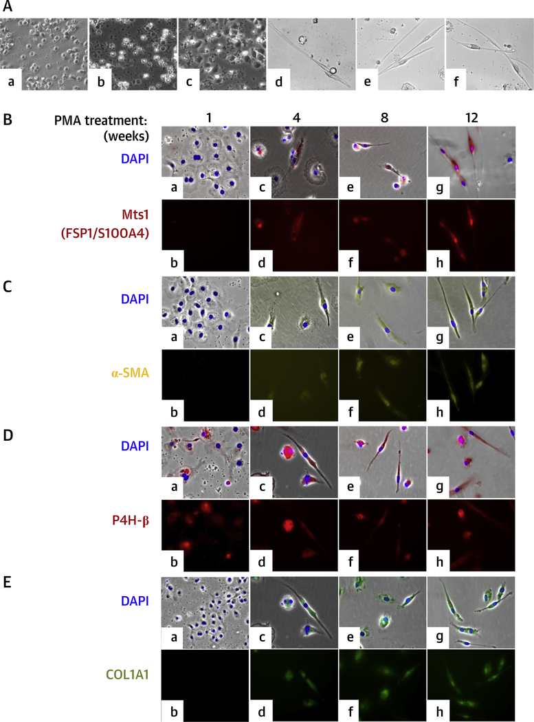
FIGURE 1. Differentiation of Human CD14+ Peripheral Blood Monocytes Into Macrophages and Expression of FSP1, α-SMA, P4H-β, and COL1A1 in Macrophages Differentiated From Human CD14+ Peripheral Blood Monocytes After PMA Treatment.
(A) Differentiation of human CD14+ peripheral blood monocytes into macrophages. (a) Freshly isolated monocytes. (b) Untreated 1-day cultured monocytes. (c) One-day PMA-treated monocytes. (d) PMA activated monocytes after 12 weeks. (e) PMA-activated and aldosterone-treated monocytes after 12 weeks. (f) PMA-activated and angiotensin II—treated monocytes after 12 weeks. Cells were visualized by phase-contrast microscopy. (B to F) Expression of FSP1, α-SMA, P4H-β, and COL1A1 in macrophages differentiated from human CD14+ peripheral blood monocytes after PMA treatment. Immunofluorescence staining with(B) Mts1 (FSP1/S100A4) mouse monoclonal antibody followed by Alexa Fluor 594 goat antimouse secondary antibody (red); (C) Cy3-conjugated α-SMA antibody (yellow); (D) P4H-β antibody followed by Alexa Fluor 594 goat antimouse secondary antibody (red); (E) FITC-conjugated antihuman COL1A1 antibody (green) of PMA-treated cells. Nuclei were stained with 4,6-diamidino-2-phenylindole (DAPI) (blue). (a and b) 1 week; (c and d) 4 weeks; (e and f) 8 weeks; and (g and h) 12 weeks after PMA activation. Cells were visualized in a Leica DMIRE2 fluorescence microscope. (a, c, e, g) Brightfield phase-contrast images merged with fluorescence images (red/yellow/green) and nuclei stained with DAPI (blue). Magnification ×200. COL1A1 = type I collagen; FSP1/S100A4 = fibroblast-specific protein-1; MI = myocardial infarction; P4H = prolyl-4-hydroxylase; SMA = smooth muscle actin.

