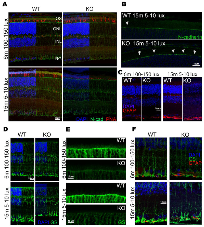
Figure 6.

Sphk1 KO mouse retina shows a loss of N-cadherin at the OLM. A) Retinas from WT and KO mice raised in both the 5-10 lux and 100-150 lux light conditions were stained for the cone outer sheath with peanut agglutinin (PNA, red) and show no change between WT and KO mice. Retinas from both WT and KO mice were also stained for N-Cadherin. Gaps in N-cadherin coverage are seen in KO retinas at the OLM. B) Magnification (2× zoom) of N-cadherin staining at the OLM in 15-month-old mice raised at 5-10 lux. There is a significant loss of the normally continuous coverage of N-cadherin (white arrowheads) at the OLM in KO mice. C) Retinas stained for glial fibrillary acidic protein (GFAP) to detect glial activation of Müller cells show that they do not become activated, and thus are not the reason for the loss of the OLM. D) Müller glia stained for glutamine synthetase (GS) show no overall loss of Müller glia cell numbers, but a loss of staining at the apical side can be detected. E) 2× magnification of the apical Müller feet that make up the OLM shows a loss of staining and villous structures that reach into the inner segments in Sphk1 KO mice. F) Basal end feet of the Müller glia that make up the inner limiting membrane remain unaffected in the KO mouse retina. Representative images from n=3 mice.
