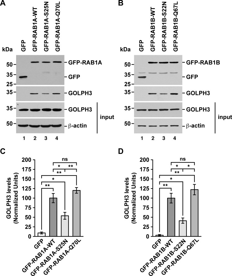
Fig 5. GFP-Trap analysis of the interaction of endogenous GOLPH3 with either RAB1A or RAB1B.
(A-B) Soluble protein extracts from human H4 neuroglioma cells transiently expressing the indicated variants of GFP-RAB1A (A) or of GFP-RAB1B (B) were subjected to the GFP-Trap assay. Samples of the GFP traps (upper two panels) and of the protein extracts used in the pulldowns (lower two panels; input) were processed by SDS-PAGE followed by immunoblotting. Immunoblottings were carried out using antibodies to detect the proteins indicated on the right. Antibody to GFP was used to detect GFP (used as GFP-Trap control) and the GFP-tagged RAB1A and RAB1B variants. The position of molecular mass markers is indicated on the left. (C-D) Densitometry quantification of the amount of GOLPH3 pulled down as shown in the third panel of A and B. The immunoblot signal of anti-β-actin was used as loading control. Bar represents the mean ± standard deviation (n = 3 independent experiments). * P < 0.05; ** P < 0.01; ns, not statistically significant.

