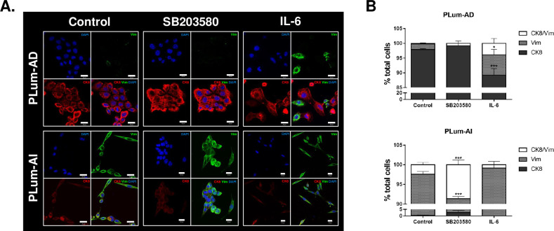
Fig 2. Immunofluorescence characterization of PLum-AD and PLum-AI cells upon treatment with SB203580 (10 μM) and IL-6 (10 ng/mL).
(A) Representative immunofluorescent images of PLum-AD and PLum-AI cells stained for EMT markers (epithelial CK8 and mesenchymal VIM) and the nuclear counterstain DAPI are shown. Scale bars = 20 μm. (B) Quantification of the percentage of cell counts expressing double positive CK8/Vim, Vim alone or CK8 alone. Data are expressed as the percentage of positively stained cells for each marker with respect to the total number of cells. Data represent an average of three independent experiments and are reported as mean ± SEM (p<0.001, Two-way ANOVA; *P<0.05; ***P<0.001; different treatment conditions compared to the control for each marker and for each cell line, Bonferroni’s multiple comparison’s test).

