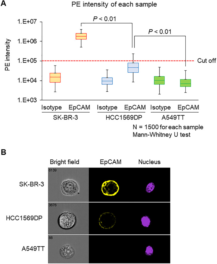
Fig 2. Evaluation of EpCAM expression level.
Each cell line was stained with a PE-anti-EpCAM antibody or an isotype control antibody. Each sample contained 1500 measured cells. (A) Intensity of PE fluorescent signals. Data are presented as a box-whisker plot. The lower whisker indicates the 2.5th percentile, and the upper whisker indicates the 99.5th percentile. The fluorescence signal cutoff of PE was set so that the positive rate of isotype control in all three cell lines was 0%. Data were analyzed using the Mann–Whitney U-test. (B) Immunostaining images obtained by imaging flow cytometry. https://doi.org/10.6084/m9.figshare.12433157.v2.

