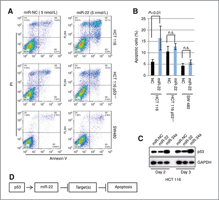
Figure 2.
A, fluorescence-activated cell sorting (FACS) analysis. HCT 116, HCT 116-p53−/−, and SW480 were transfected with 5 nmol/L of miR-22 or miR-NC, incubated for 3 days, and subjected to FACS analysis. B, quantification of apoptotic cells. Apoptotic cells were quantified by using 4 independent FACS experiments. Data indicate the mean value with SD. Statistical analysis was carried out by t test. C, p53 is not activated by miR-22. Cells, transfected with miR-22 or miR-NC, were incubated for 2 or 3 days, and subjected to immunoblotting. D, hypothesis of miR-22 function in the p53 network.

