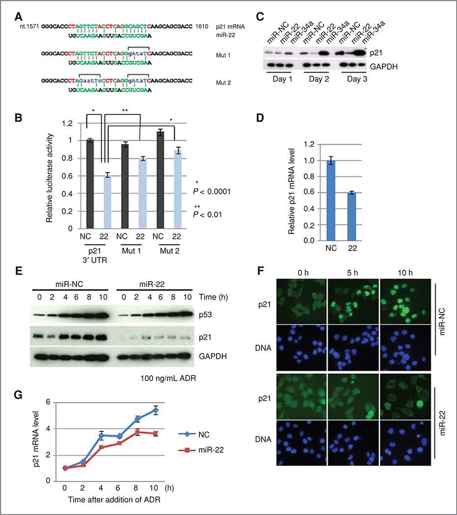
Figure 4.
A. sequence alignment of miR-22 and the 3’ UTR of p21 mRNA is indicated at the top. Mutant sequences used for the reporter gene assay are listed (Mut 1 and Mut 2). B, reporter gene assay. Error bar indicates SD (n = 6). C, expression level of p21 protein in the presence of miR-22. MiR-34a was used as positive control. D, expression levels of p21 mRNA in the presence of miR-22. Cells were transfected with miR-22, and incubated for 3 days. The relative expression levels of p21 mRNA were quantified by TaqMan assay. E, effect of miR-22 on the activation of p21 expression after exposure to ADR. HCT 116 cells were transfected with 5 nmol/L of either miR-22 or miR-NC and incubated for 48 hours, and further incubated in the presence of ADR for the indicated times. F, indirect immunocytochemistry. Cells were transfected as described above, and incubated in the presence of ADR for the indicated times. Cells were subjected to immunostaining. G, activation of p21 expression in the presence or absence of miR-22 after exposure to ADR. Cells were prepared as described in (E), and total RNAs were prepared from each time point. Relative expression levels of p21 mRNA were quantified by TaqMan assay.

