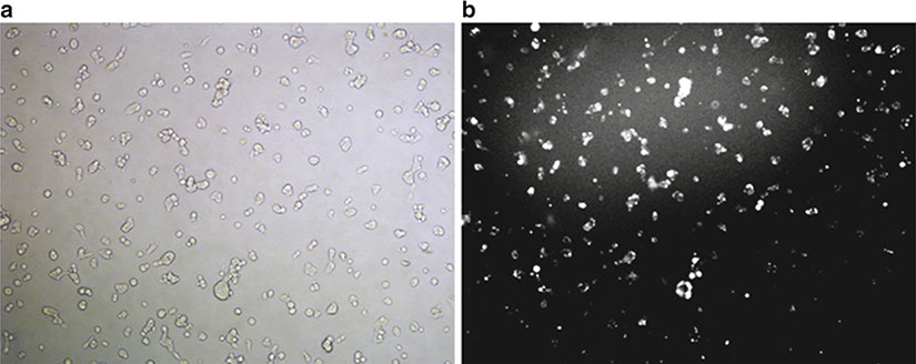
Fig. 1.
Visualization of siRNA localization after a typical nucleofection reaction. HEK-293 cells were subject to nucleofection with fluorescent siRNA (siRNA-GLO Red, Thermo Scientific, Lafayette CO). After 24 h, the cells were visualized and bright field (a) and fluorescent (b) images were captured. The fluorescence reveals efficient delivery of siRNA, predominantly localized to the cytosol and nucleus.

