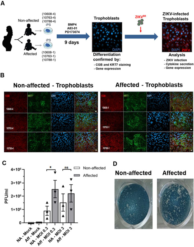
Fig 1. Experimental design and ZIKVBR infection in hiPSC-derived trophoblasts.
(A). Schematic: generation of trophoblasts from congenital Zika syndrome affected and non-affected discordant twins’ hiPSCs followed by ZIKVBR infection and analysis. Silhouettes are courtesy of www.vecteezy.com (mother) and Yulia Ryabokon (babies). (B). Immunofluorescence for CGB (β-CG, human chorionic gonadotropin β) and KRT7 (CK7, cytokeratin 7, a pan trophoblast marker) in hiPSC-derived trophoblasts. Scale bar: 20 μm. (C). ZIKV PFU/mL in trophoblasts’ supernatant at MOI = 0.3 and MOI = 3; mean ± SEM of the three twins; *p < 0.05; Student’s t test. (D). Representative plaque forming assay wells with stained VERO cells exposed to ZIKV collected at 96 hpi from the culture supernatants of affected or non-affected #10608 twins’ hiPSC-derived trophoblasts infected at a MOI of 0.3.

