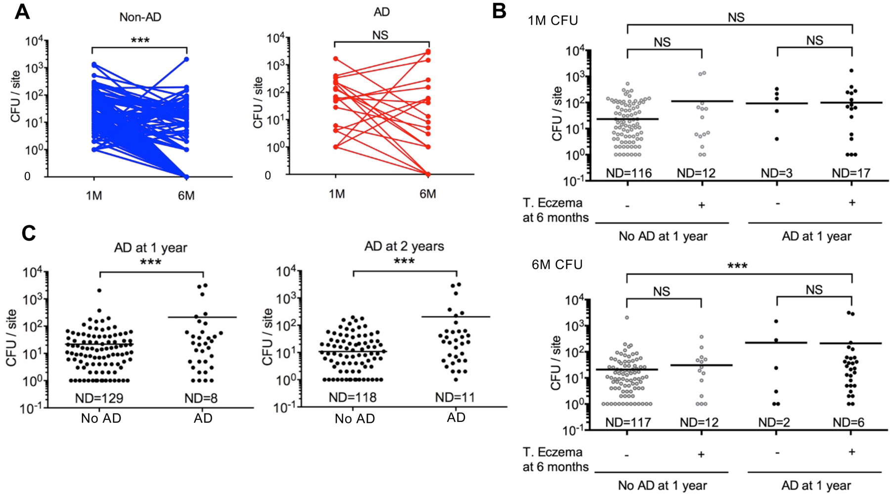
Fig. 1. Increased S. aureus colonization in infant cheek skin at 6 months is associated with AD development.

A, S. aureus colony-forming units (CFU) in the cheek skin (CFU per site) of infants at 1-month (1M) and 6-month (6M) of age. Infants were colonized with S. aureus at 1 month. Lines indicate samples from the same infant. Blue indicates infants who did not develop AD (No AD), red indicates infants who developed AD (AD). ***p<0.001, Two-tailed Wilcoxon signed rank test. B, Each dot represents the number of CFU per site (cheek skin) from an infant at 1 month (1M) or 6 months (6M). Infants who developed and did not develop AD at 1 year were subgrouped by the presence of unclassified transient eczema (T. eczema) at 6 months. Transient eczema was defined by the presence of eczema symptoms for less than 2 months. ***p<0.001, Kruskal-Wallis test with Dunn’s post hoc test. . C, S. aureus CFUs in the cheek skin. Each dot represents results from an infant at 6 months. Number of CFUs/per site in infants who did or did not develop AD at 1 year and 2 years of age. ***p<0.001, Mann-Whitney test. ND, not detected; NS, not significant..
