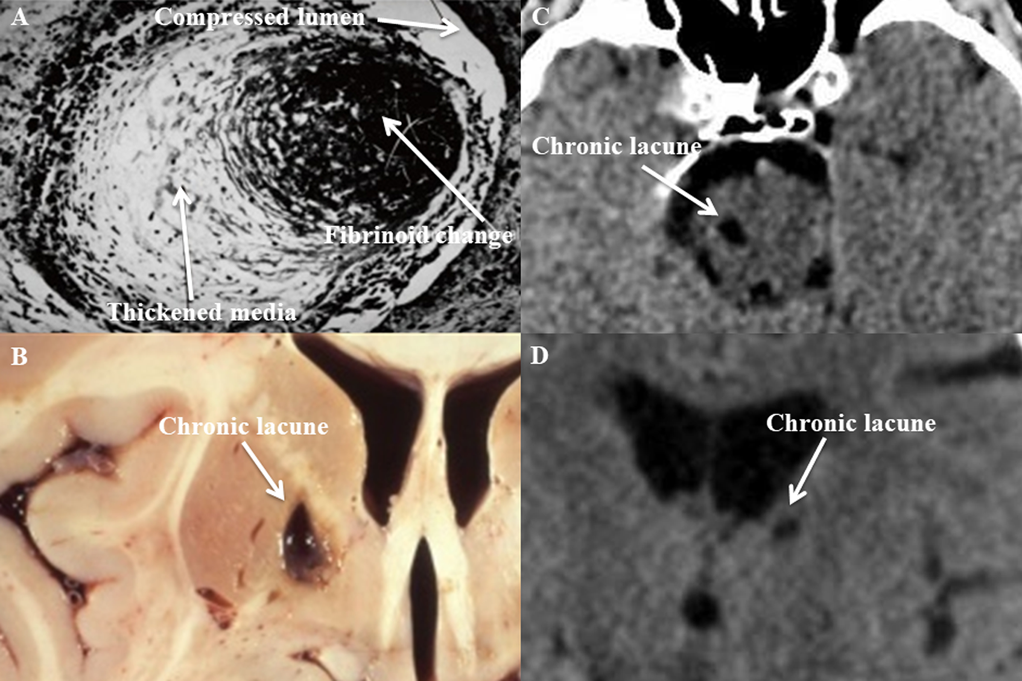
Figure 1. Cerebral small vessel disease (cSVD) histopathological section, lacunar stroke gross section, and lacunar stroke computed tomography (CT) images.

Photomicrograph of cSVD affecting a small penetrating artery (A), showing thickened media and fibrinoid necrosis (Gift from Dr. C. Miller Fisher). Photograph of a cavity secondary to a chronic lacune in the medial basal ganglia (B) found at autopsy.2 CT images showing hypodensities secondary to chronic lacunes involving the right paramedian pons (C) and left thalamus (D).
