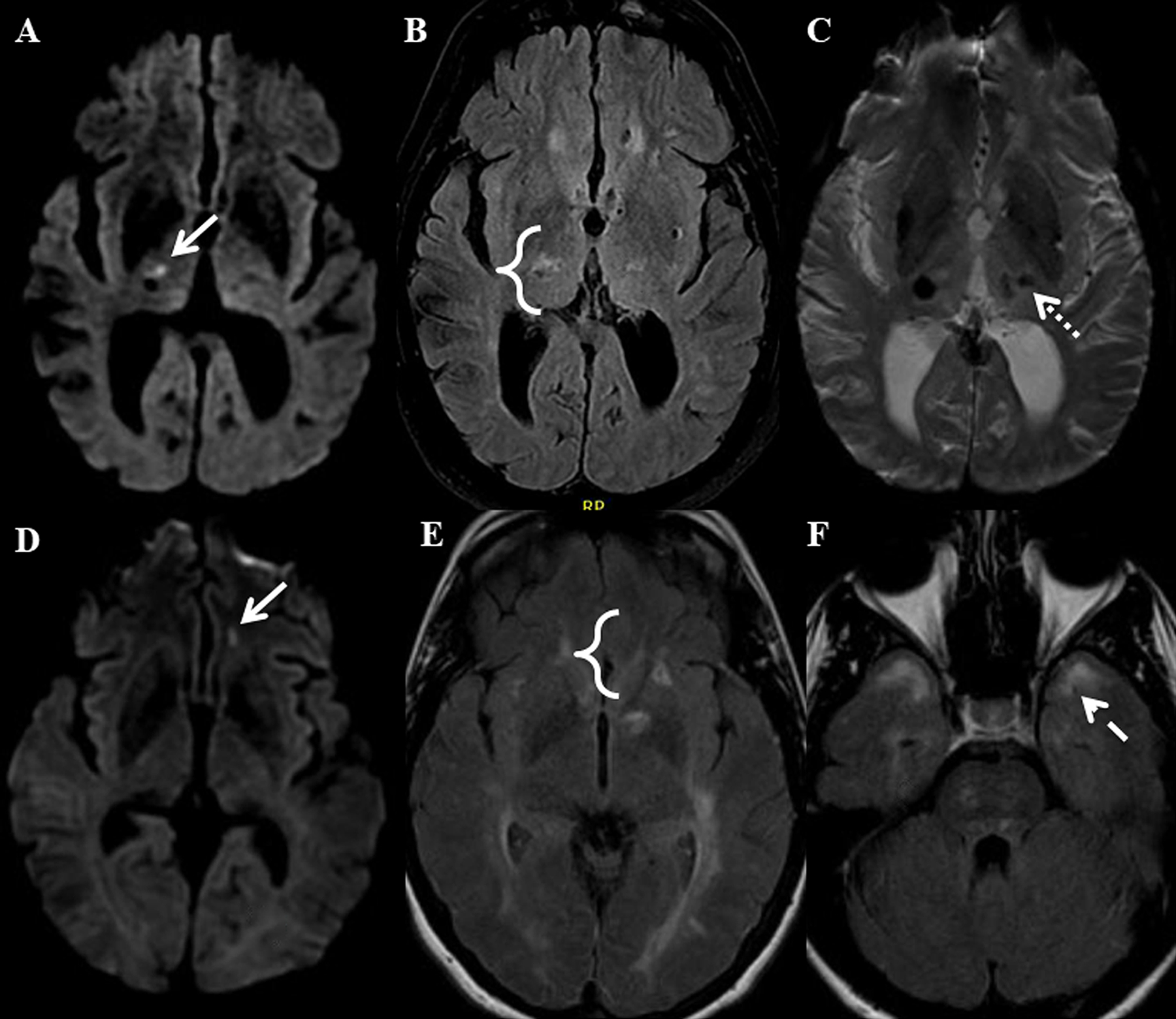
Figure 3. Lacunar stroke (LS) and white matter hyperintensity (WMH) proximity.

Panels A-C show the brain of a patient with severe sporadic hypertension-related cerebral small vessel disease (cSVD). An acute LS (arrow) is shown in the right thalamus on DWI imaging (A). FLAIR imaging (B) shows there is an adjacent WMH (bracket). SWI imaging (C) shows there is also a microhemorrhage in the left thalamus (dotted arrow). Panels D-F show the brain of a patient with CADASIL. An acute LS (arrow) is shown in the left orbitofrontal white matter on DWI imaging (D). FLAIR imaging (E) shows there is an adjacent WMH (bracket). WMH in the anterior temporal lobe (dashed arrow) is also present on FLAIR imaging (F).
