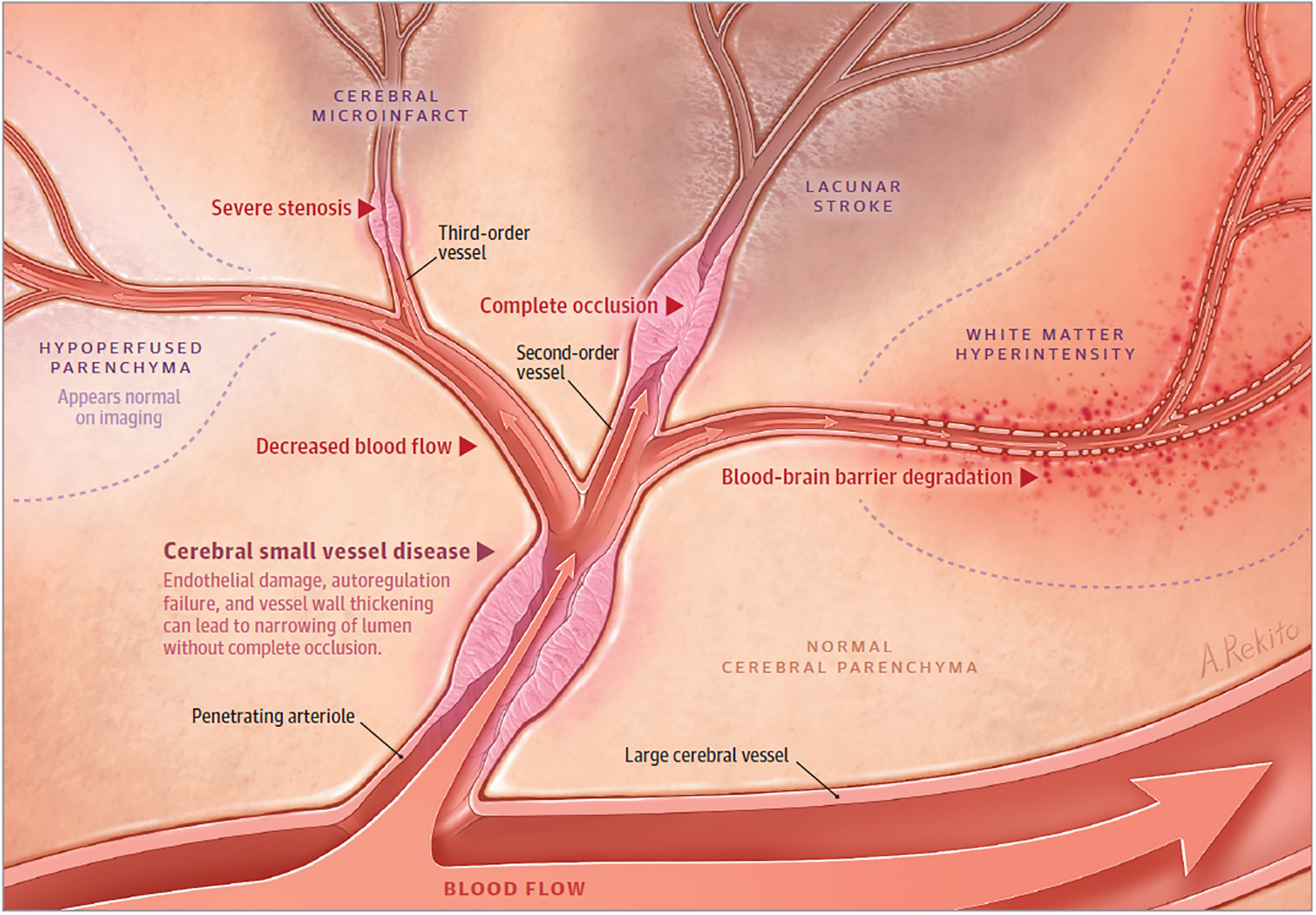
Figure 4.

Originating from a large vessel, a penetrating arteriole is shown with narrowing and impaired autoregulation from cerebral small vessel disease. Downstream, there is hypoperfused parenchyma that appears normal on imaging. In the setting of occluded cerebral small vessel disease in a second-order vessel, lacunar stroke occurs. In higher-order vessels with occluded cerebral small vessel disease, smaller lacunar strokes and cerebral microinfarcts occur. Areas of blood-brain barrier degradation with decreased cerebral blood flow result in white matter hyperintensity. Lacunar strokes align with penetrating vessels, but they also have a predilection to form at the edge of white matter hyperintensities. Hypoperfused parenchyma can progress to lacunar stroke and white matter hyperintensity, although these sequelae of cerebral small vessel disease can also improve over time. The dynamic nature of lacunar stroke likely results from a balance of blood flow changes, focal cerebral small vessel disease changes, and parenchymal repair processes.
