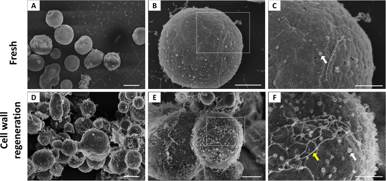
FIG 1.
Morphological aspects of freshly prepared and cell wall-regenerating protoplasts. Fresh protoplasts (A to C) and cell wall-regenerating cells (D to F) are shown under conditions of increased magnification by SEM. Panels C and F represent magnified views of the boxed areas in panels B and E, respectively. The magnified views suggested the occurrence of outer particles with properties compatible with EVs (white arrows). Under cell wall-regenerating conditions, a fibril-like network was more abundantly detected (yellow arrow). Scale bars represent 5 μm in panels A and D, 2 μm in panels B and E, and 1 μm in panels C and F. At least 50 cells were analyzed, and the results are representative of at least two independent experiments producing similar morphological profiles. Similar analyses using superresolution SEM produced similar results (data not shown).

