FIG 2.
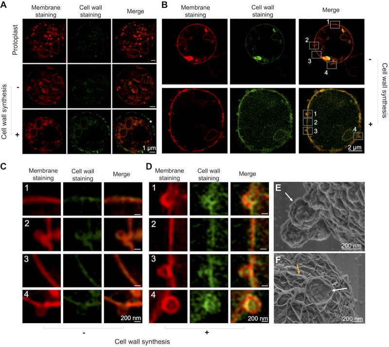
Membrane projections in A. fumigatus protoplasts. (A) Freshly purified protoplasts were stained with DiI, a lipophilic dye (red fluorescence). Cell wall staining with an anti-glucan antibody was at the background levels. Similar results were observed for protoplasts incubated under nonregenerating conditions. During cell wall regeneration (2 h), glucan staining (green fluorescence) was abundant at the cell surface (asterisk). (B) Detailed analysis of nonregenerating and cell wall-regenerating cells revealed an association between glucan staining and outer membrane projections only in cell wall-regenerating protoplasts (90 min of incubation). (C and D) Enhanced views of the boxed areas (numbered 1 to 4) of fungal cells in the absence of cell wall synthesis and under cell wall-regenerating conditions, respectively. (E and F) A detailed view of the surface of protoplasts provided by superresolution SEM confirmed the occurrence of outer particles (white arrows) budding from the plasma membrane in nonregenerating protoplasts (E) and regenerating (2 h) protoplasts (F). Fibrillar material closely associated with the outer membrane projection was uniquely detected during cell wall regeneration (F, yellow arrow). At least 10 cells were analyzed, and the results are representative of two independent experiments producing similar morphological profiles.

