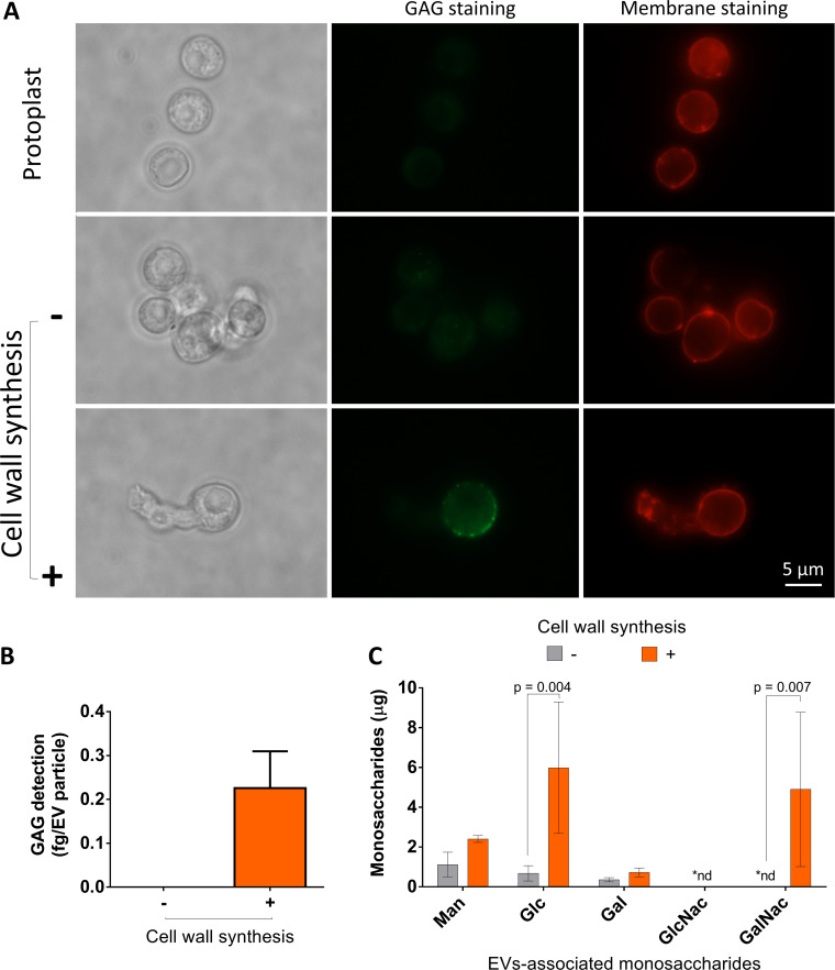
FIG 5.
Analysis of glycan synthesis during cell wall regeneration in A. fumigatus conidial protoplasts. (A) Membrane and GAG staining in A. fumigatus protoplasts. All cells were efficiently stained with DiI (red fluorescence). During cell wall synthesis (2 h), GAG was detected in association with the fungal surface. The scale bar corresponds to 5 μm. (B) Serological detection of GAG (ELISA) in EVs obtained from protoplasts. Positive reactions with a GAG-binding antibody were observed only in EVs obtained during cell wall synthesis. (C) Gas chromatography-mass spectrometry (GC-MS) analysis of sugar units of EVs. In agreement with an involvement of EVs in cell wall synthesis, GalNAC (a GAG component) was observed only in EVs obtained from protoplasts during cell wall regeneration. The increased detection of Glc during cell wall synthesis (2 h of germination) is consistent with the presence of EV-associated glucans. The results are representative of two independent replicates producing similar profiles.

