
Figure 1.
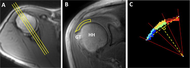
Methodology for segmentation. (A) Using an axial localizer image, three central-most coronal-oblique images relative to the humeral head were identified (yellow lines). (B) Coronal-oblique image demonstrates manually segmented RCT from the center of the humeral head to the greater tuberosity attachment. (C) For each manually segmented region on each slice, six subsegments of equal size were automatically divided based on bursal-sided (dark red, orange, and light blue) and articular-sided (red, green, and dark blue) halves as well as medial (dark red and red), middle (orange and green), and lateral thirds (light blue and blue). HH: humeral head, GT: greater tuberosity.