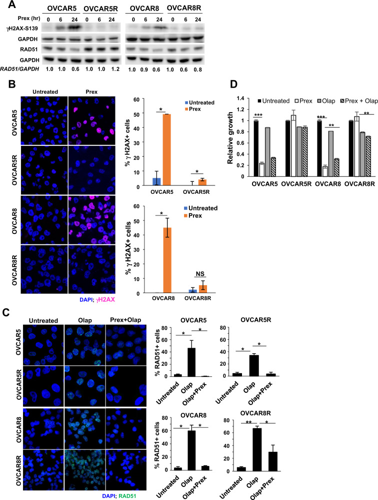
Fig. 4. Effects of CHK1i on DNA damage and CHK1 activation.
a Immunoblotting analysis of a DNA damage marker γH2AX and an HR marker RAD51 were performed in cells treated with Prex (20 nM) for 6 and 24 h. Densitometric quantifications of RAD51 normalized with GAPDH and relative ratios are shown. b Parental and PrexR cells were cultured on coverslips overnight with or without Prex (20 nM) and then fixed and stained with antibodies against γH2AX (pink) and nuclear stain DAPI (blue). Cells with >5 γH2AX foci were counted as γH2AX-positive (γH2AX+) cells. Percentage of γH2AX+ cells are plotted on the right. c Immunofluorescence staining of parental and PrexR cells for RAD51 foci (green) induced by PARPi olaparib (Olap) (20 µM) with or without Prex (10 nM). Nuclei were stained with the nuclear stain DAPI (blue). Cells with >5 RAD51 foci were counted as RAD51-positive (RAD51+) cells from three fields of on each slide and the percentage of RAD51+ cells are plotted on the right. All experiments were repeated at least thrice and representative images are shown. d XTT growth assay on parental and PrexR cells after 48 h treatment with Prex (10 nM) with or without Olap (20 µM). *P < 0.05; **P < 0.01; ***P < 0.001; NS not significant.

