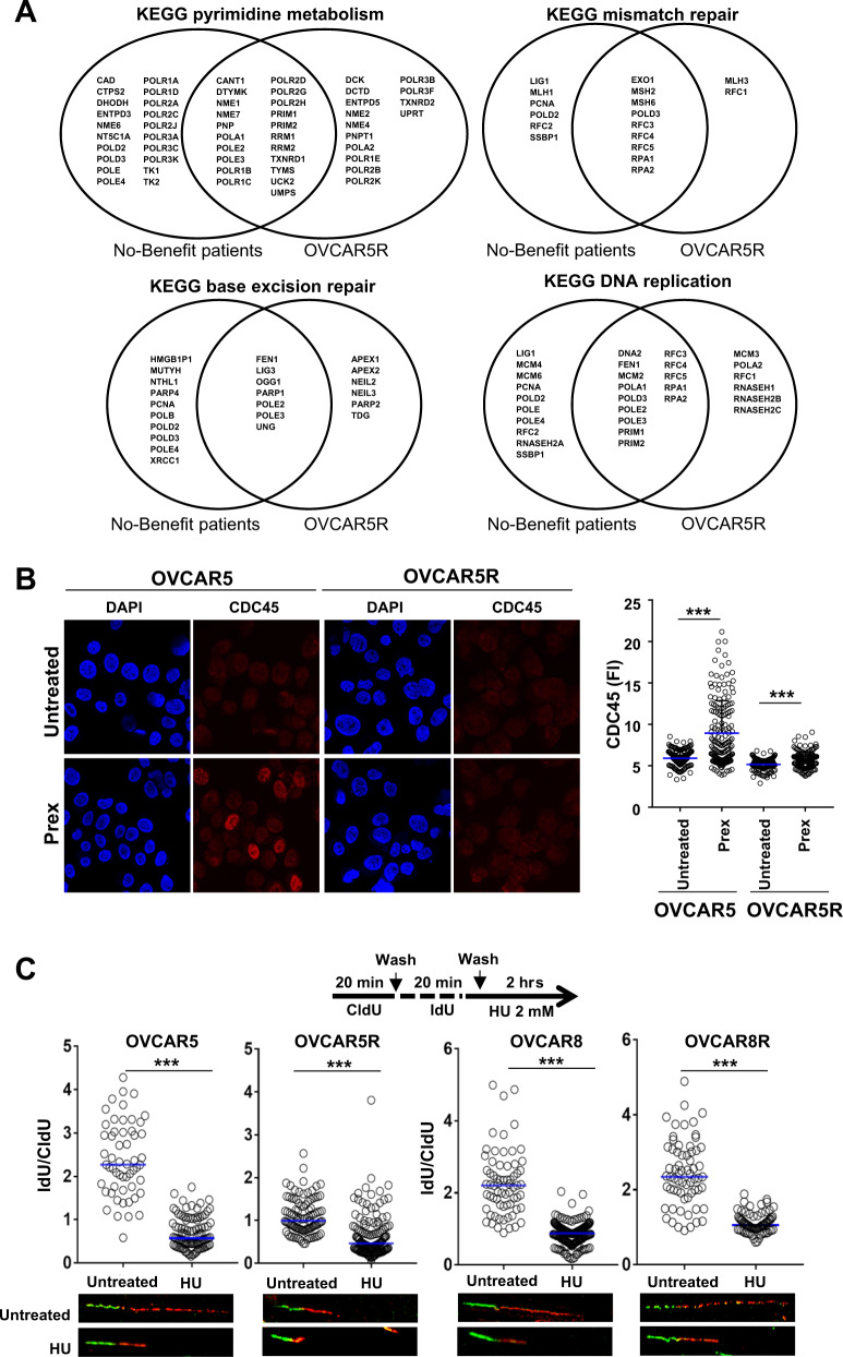
Fig. 5. Roles of the replication fork and DNA repair pathways in CHK1i resistance.
a Venn diagram plots of genes that contribute to the enrichment of gene sets as represented in Table 1. Common genes between the no-benefit patient group (n = 7) and the Prex-resistant OVCAR5R (n = 3) appear at the intersection between the two groups. b Immunofluorescence staining for DNA replisome protein CDC45 after pre-extraction was performed in OVCAR5 and OVCAR5R cells cultured overnight with or without Prex (20 nM). Fluorescence intensity (FI) for CDC45 was quantified for at least 200 cells by using ImageJ and plotted on the right. c DNA fiber assays were done on parental and PrexR cells. Cells were stained with CldU and IdU, followed by treatment with 2 mM HU for 2 h. Resection of nascent DNA strands induced by HU treatment (IdU labelled) was estimated as a ratio of IdU labelled tracts to CldU for at least 100 strands and plotted as median at 95% CI. Images of representative strands are shown below each plot. All experiments were repeated at least in triplicate and representative data is shown. All experiments were performed at least thrice. Data are shown as mean ± SD. ***P < 0.001; NS not significant.

