
Fig. 1.
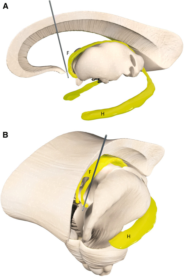
Simplified illustration of anatomical targeting for fornix deep brain stimulation in clinical studies. The fornix (F) and the hippocampus (H) are depicted in yellow. Efferent fibers of the hippocampus known as the alveus join together to form the fimbria. Beneath the splenium of the corpus callosum, the fimbria separates from the hippocampus and becomes the crus of the fornix. The left and right crura then converge to form the body of the fornix. The body of the fornix travels anteriorly and divides again near the anterior commissure. The left and right parts separate into the anterior pillars, and there is also an anterior/posterior divergence. The posterior fibers (called the postcommissural fornix) of each side continue through the hypothalamus to the mammillary bodies. The anterior fibers (precommissural fornix) end at the septal nuclei and nucleus accumbens of each hemisphere. a Sagittal view of fornix DBS electrode location. b Frontal view of fornix DBS electrode location in one hemisphere