Fig. 1.
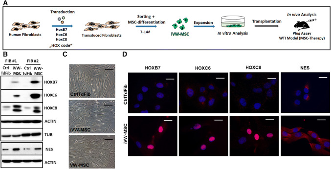
Induction of vascular wall-typical MSCs from adult human fibroblasts. a Experimental design for the induction of human MSCs. Primary human fibroblasts were transduced with a lentiviral SIN vector co-expressing the coding sequences of HOXB7, HOXC6 and HOXC8 and Turquoise2 (Cyan), which are all co-translationally separated by 2A esterase moieties [48]. Two to four days after transduction the cells were sorted for cyan fluorescence and cultured in MSC medium. Generated MSCs (iVW-MSC) were characterized 14 days after induction when cells were sufficiently expanded. b Western blot analysis of total HOXB7, HOXC6 and HOXC8 protein expression levels as well as of Nestin (NES) were performed from whole cell lysates of HOX-transduced (iVW-MSC) and control fibroblasts (CtrlTdFib) 12–14 days after isolation of transduced cells. Representative blots from two transductions of fibroblasts from two independent donors are shown. Beta-actin (ACTIN) and alpha-tubulin (TUB) were included as loading controls. #Fibroblasts derived from different healthy donors. c Representative phase contrast micrographs of cells 10–12 days after flow-cytometric sorting showed typical mesenchymal cell morphology. Ex vivo isolated hITA (human internal thoracic artery)-derived VW-MSCs were shown as control. d Sorted HOX-transduced and control fibroblasts were seeded on gelatine-coated cover-slips and HOXB7, HOXC6, HOXC8, and NES expression were detected by immunofluorescence using confocal microscopy. Representative photographs are shown

