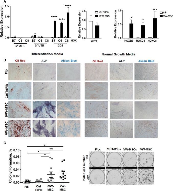
Fig. 2.
Characterization of control- and HOX-transduced fibroblasts (iVW-MSCs). a Relative amounts of transcripts of the indicated genes were determined by qRT-PCR in iVW-MSCs and control vector-transduced fibroblasts (CtrlTdFib) 12–14 days after transduction and flow-cytometry based cell isolation (biological replicates: n = 4–6 per group and gene; P by two-way ANOVA, followed by post hoc Tukey’s multiple comparisons test: ****P ≤ 0.001). For the detection of endogenous HOX gene expression, desoxy-oligonucleotide-primer pairs located in the 3′UTR as well as in the 5′UTR were used, which are not present in the retroviral expression vector containing only the coding sequence (CDS). For overall detection of retroviral vector expression, the woodchuck hepatitis virus posttranscriptional regulatory element (wPRE) was used. Differential HOX gene expression levels (CDS) in ex vivo isolated hITA (human internal thoracic artery)-derived VW-MSCs as compared to primary (non-transduced) fibroblasts (Fib) were included as positive and negative control. (biological replicates: n = 4–6 per group and gene; P by two-way ANOVA, followed by post hoc Tukey’s multiple comparisons test: *P ≤ 0.05; ***P ≤ 0.005). Relative transcript levels of analyzed genes were normalized to beta-actin mRNA (set as 1). b Verification of induced conversion into MSCs. FACS-purified iVW-MSCs and control fibroblasts were differentiated into adipocytes, osteocytes and chondrocytes, in vitro. Differentiation was observed within 14 days after induction of differentiation (DM) as shown by Oil red staining for detecting lipid droplets (red) in adipocytes, by histochemical NBT/BCIP staining for detecting alkaline phosphatase activity (ALP, black-purple) in osteocytes, or Alcian Blue staining (blue) for detecting acidic polysaccharides such as glycosaminoglycans in (e.g. the cartilage-specific proteoglycan aggrecan) in chondrocytes. Representative photographs are shown (biological replicates: n = 3–4). Magnification × 400. As control, respective cells were cultured in normal growth media (NGM). c CFU Assay. Control-transduced fibroblasts and iVW-MSCs were plated at low densities (100–1000 cells/well) in plastic culture dishes and subsequently cultured for 10 days. The lack of the colony-forming potential of primary fibroblasts as compared to hITA-derived VW-MSCs was confirmed by plating respective cells. Coomassie Brilliant Blue stained colonies were counted and the surviving fraction (colony formation) was calculated. P by two-way ANOVA, followed by post hoc Tukey’s multiple comparisons test: *P ≤ 0.05, **P ≤ 0.01 (biological replicates: n = 8–10 for each group)

