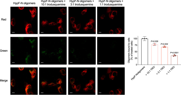
Fig. 2. Trodusquemine reduces the membrane binding affinity of HypF-N oligomers to cultured human neuroblastoma cells.
Representative confocal scanning microscopy images of the apical sections of SH-SY5Y cells treated for 15 min at 37 °C with HypF-N oligomers (6 µM) in the absence (black) or presence of 10:1, 3:1, and 1:1 ratios of HypF-N to trodusquemine (light to dark red). Red and green fluorescence indicates the cell membranes and the oligomers, respectively. The percentage of colocalization between membranes and HypF-N oligomers are shown. In all, 50–60 cells were analyzed per condition. Bars indicate the mean ± s.e.m. of n = 3 biologically independent experiments (dots). Statistical analyses were carried out by one-way ANOVA relative to cells treated with oligomers in the absence of trodusquemine. Scale bars, 10 µm.

