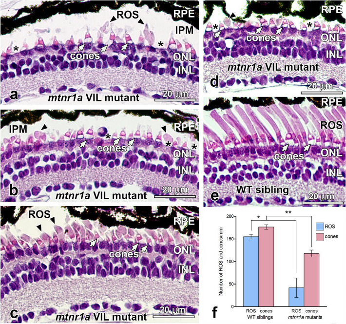
Figure 3.
Photoreceptors degenerate in stage 50–52 (35-day-old) heterozygous mtnr1a VIL mutant tadpoles. (a–d) H&E-stained paraffin sections of the central retina of four different stage 50–52 mtnr1a VIL mutant tadpoles display rod loss and/or dystrophy. Retinas of mutant tadpoles have normal appearing cones (white arrows), but severely dystrophic rod outer segments (ROS; black arrowheads). Note the frequent absence of rod inner segments (asterisks). (b) A presumptive leukocyte is in the area of the interphotoreceptor matrix (IPM) at the center of the panel. (c,d) Some ROS persist in the mutant central retinas, but are much shorter than the ROS of WT sibling retinas (e), and exhibit some dystrophy. (e) Retinas of WT siblings exhibit healthy rod and cone photoreceptors. (f) Quantitative analysis of ROS and cones in WT vs VIL central retinas. The average number of ROS (blue bars) or cones (red bars) in 6–12 different retinal sections from four different animals (a–d) in each group (i.e., n = 4) were analyzed for statistical significance by an unpaired two-tailed t-test. Error bars indicate standard error of the mean (SEM). In mutant retinas, the number of ROS declined by 73% (*; p = 0.0022), and cones declined by 33% (**; p = 0.0008) as compared to WT. RPE retinal pigment epithelium, ROS rod outer segments, ONL outer nuclear layer, INL inner nuclear layer, IPM interphotoreceptor matrix, cone inner segments; white arrows, ROS; black arrowheads, inner segment layer; asterisks. Magnification bars = 20 µm.

