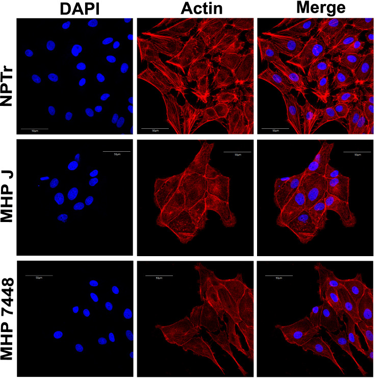
Figure 3.
Organization of actin fibers in cells infected with M. hyopneumoniae. Results of the immunofluorescence microscopy analysis indicating the reduction and change in the pattern of actin stress fibers in infected cells. Eukaryotic cell actin was labeled with phalloidin (red) and nuclei were stained with DAPI (blue). Both attenuated (J) and virulent (7448) strains of M. hyopneumoniae altered the organization and abundance of actin fibers after 24 h of infection, however this effect can already be seen after 1 h of incubation (not shown). NPTr - uninfected cells. MHP J - NPTr cells infected with M. hyopneumoniae strain J. MHP 7448 - NPTr cells infected with M. hyopneumoniae strain 7448. Scale bars:

