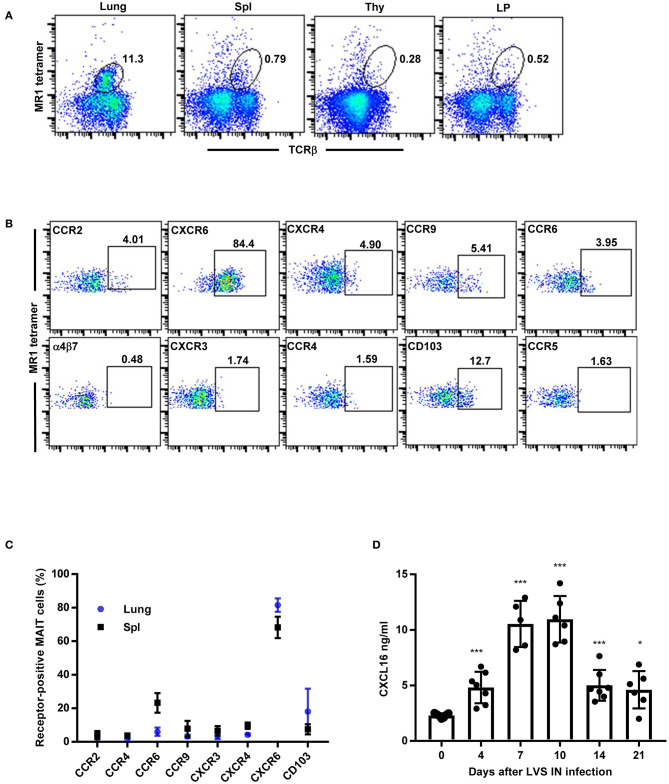
Figure 2.
MAIT cells in the lungs of WT mice predominantly express the chemokine receptor CXCR6 during F. tularensis LVS intranasal infection. (A) Flow cytometry analysis of MAIT cells in the lungs, Spl (spleen), Thy (thymus), and LP (lamina propria) of LVS-infected WT mice on day 14 after LVS infection, showing reactivity to MR1-5-OP-RU tetramer in total lung cells. (B) Representative flow cytometry dot plots depicting expression of different chemokine receptors on MAIT cells in LVS-infected WT mice lungs (day 14 after infection). Plots show live 5-OP-RU MR1 tetramer+ TCRβ+ cells. (C) A graphical representation of the percentage of MAIT cells positive for the indicated chemokine receptors in the lungs and spleens (Spl) of LVS-infected WT mice on day 14 after infection. (D) The levels of CXCL16 in lung homogenates of naïve and LVS-infected WT mice at the indicated time points (*P < 0.05; ***P < 0.001 as compared to naïve Day 0 mice). Data are presented as the mean ± SEM (n = 5–6) and are representative of two independent experiments.

