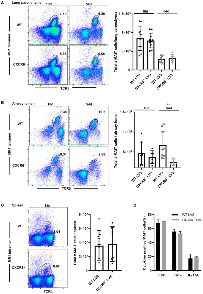
Figure 3.
CXCR6 is not required for MAIT cell accumulation in the lung parenchyma or spleen during F. tularensis LVS intranasal infection but contributes to long-term retention in the airway lumen (BAL). (A) Representative flow cytometry dot plots showing the percentage of MAIT cells in the lung parenchyma of WT and CXCR6−/− mice on days 10 and 54 after LVS IN infection. MAIT cells are gated on live MR1-5-OP-RU tetramer+ TCRβ+ cells in the lungs. A graphical representation of the number of MAIT cells in the lung parenchyma is shown. (B) Representative flow cytometry dot plots of the percentage of MAIT cells in the airway lumen (BAL) of WT and CXCR6−/− mice on days 10 and 54 after LVS IN infection. MAIT cells are gated on live MR1-5-OP-RU tetramer+ TCRβ+ cells in the lungs. A graphical representation of the number of MAIT cells in the airway lumen is shown (**P < 0.01 as compared to WT mice). (C) Representative flow cytometry dot plots of the percentage of MAIT cells in the spleen of WT and CXCR6−/− mice on day 14 after LVS IN infection. MAIT cells are gated on live MR1-5-OP-RU tetramer+ TCRβ+ cells in the spleen. A graphical representation of the number of MAIT cells in the spleen is shown. (D) Lungs harvested from LVS-infected WT and CXCR6−/− mice on day 14 after infection were stimulated in vitro in the presence of PMA and ionomycin. A graphical representation of the percentage of MAIT cells positive for IFN-γ, TNF, and IL-17A is shown. Data are presented as the mean ± SEM (n = 5–7) and is representative of at least two independent experiments.

