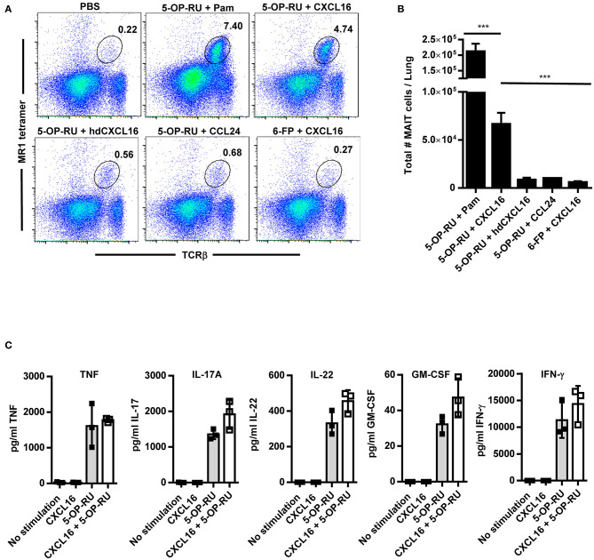
Figure 5.
Intranasal instillation of CXCL16 and 5-OP-RU induces MAIT cell accumulation in the lungs in the absence of infection. Naïve WT mice were intranasally administered 5-OP-RU or control Ac-6-FP (6-FP) with one of the chemokines shown on days 1, 2, and 3. Mice given 5-OP-RU + Pam were intranasally administered 5-OP-RU and Pam on day 1, and 5-OP-RU alone on days 2 and 3. On day 7, the lungs were harvested for flow cytometry analysis. “PBS” mice received only PBS at the indicated time points. (A) Representative flow cytometry dot plots showing 5-OP-RU MR1 tetramer+ TCRβ+ MAIT cells in naïve WT mice treated as indicated. Total live singlet lung cells for individual mice are shown. (B) A graphical representation of the number of MAIT cells in the lungs of mice on day 7 (***P < 0.001). (C) Vα19iTgMR1+/+ transgenic murine MAIT cells were co-cultured with uninfected macrophages, recombinant CXCL16, and 5-OP-RU, and cytokine production was measured after 16 h. “No stimulation” = uninfected macrophages and MAIT cells. hd CXCL16 = heat-denatured CXCL16. Pam = Pam2CSK4. Data are presented as the mean ± SEM (n = 3) and is representative of three independent experiments.

