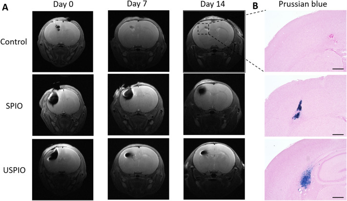
Figure 5.
In vivo results: (A) T2-weighted MRI of the control (upper, n = 1), SPIO (middle, n = 4), and USPIO (lower, n = 4) animal groups at 0, 7, and 14 days after injection of UC-MSCs. (B) Micrographs of the low-signal-intensity regions observed on MRI at day 14. SPIO and USPIO deposits are demonstrated in the left hemisphere by Prussian-blue staining. Scale bars, 500 μm. UCMSCs umbilical cord-derived mesenchymal stem cells, SPIO superparamagnetic iron oxide, USPIO ultrasmall superparamagnetic iron oxide, MRI magnetic resonance imaging.

