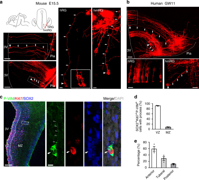
Fig. 1. Progenitor composition in the developing mammalian hypothalamus.
a, b The mouse and human hypothalamic VZ and MZ are populated with radial glia and monopolar radial glia-like cells. The morphology of hypothalamic progenitors was labeled with DiI crystals on the pial surface. The insert shows dye diffusion along radial fibers (arrow) from the hypothalamic pia terminating at distinct cell bodies (arrowhead) in the MZ or along the radial fibers traversing to the radial glia cells in the VZ at E15.5 in mice (a, n = 6 independent experiments) and GW11 in humans (b, n = 3 independent experiments). Dashed lines indicate the surface of the third ventricle. Dotted lines indicate the pial surface. 3V, the third ventricle. Scale bars, 100 µm (left, top and bottom), 20 µm (middle), 10 µm (right) in a, 50 µm (top) and 20 µm (bottom) in b. c P-VIM+ (green) hmRG cells in the GW10 human hypothalamus were stained for SOX2 (blue) and Ki67 (red). High-magnification images of the representative outlined cell are shown in the right panels. Arrows indicate hmRG cells that co-expressed Ki67 (red) and SOX2 (blue). Open arrowheads indicate the basal processes. Scale bars, 200 μm (left) and 10 μm (right). d Quantification of the percentage of RG cells identified by SOX2+Ki67+P-VIM+ immunostaining in the VZ and MZ (n = 3 independent experiments). e Quantification of the percentage of hmRG cells (SOX2+Ki67+P-VIM+) in the MZ from the anterior to posterior hypothalamus (n = 3 independent experiments). Data are presented as mean values ± SEM. Source data are supplied as a Source Data file.

