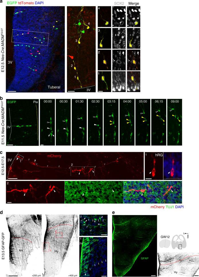
Fig. 3. Early hypothalamic neurogenesis occurs in radial columns.
a Labeling of radial arrays of cells (n = 3 independent experiments) in the developing mouse hypothalamus using MADM. A high-magnification image of the radially aligned clones is shown. The cellular composition of clones was identified by staining for the radial glial progenitor marker SOX2. Dashed lines indicate the VZ surface. White dotted lines indicate cell bodies. Right, magnified images of RG cells (area 1) and progeny (areas 2–4). Scale bars, 50 μm (left), 20 μm (middle) and 10 μm (right). b Time-lapse imaging of a MADM-labeled radially aligned clone (n = 3 independent experiments). Arrows indicate RG cells. White arrowheads indicate hmRG cells. Yellow arrowheads indicate daughter cells. Open arrowheads indicate migrating progeny. Time stamp, h:min. Scale bars, 50 μm (left) and 20 μm (right). c Representative mCherry-retrovirus-labeled progeny of a radial glial cell 5 days after the injection at E12.5 (n = 3 independent experiments). mCherry-positive hRG cell (box 1) located on the surface of the 3V. The mCherry-expressing progeny (box 2) in the MZ were positive for the neuronal marker TUJ1 (green). Arrows indicate the hRG cells. Arrowheads indicate daughter cells. Open arrowheads indicate basal processes. Dashed lines indicate the surface of the 3V. Scale bars, 50 µm (top left) and 10 µm (top right and bottom). d Spatial distribution of GFAP+ radial fibers at E13.5 (n = 4 independent experiments). Arrows indicate GFAP-expressing hRG cells (box 1) aligning at the surface of the third ventricle, and arrowheads indicate cells (box 2) migrating along radial fibers (open arrowheads). We changed the green GFP to black–white version to make it more visible. DNA, blue. Red dashed lines define the presumptive hypothalamus. Scale bars, 200 µm (left) and 20 µm (right). e GFAP expression in the developing human hypothalamus (n = 3 independent experiments). Scale bars, 400 µm (left and right).

