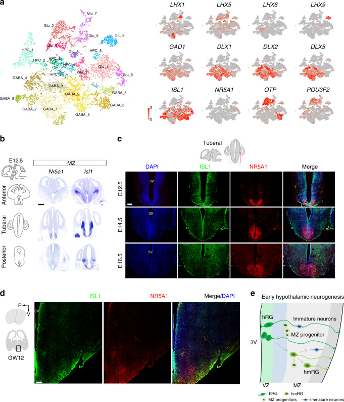
Fig. 6. Specific regional transcription factors in the developing mammalian hypothalamus.
at-SNE plot displaying well-known markers for neuron subpopulations (gray, no expression; red, relative expression). b In situ hybridization of Isl1 and Nr5a1 at E12.5 hypothalamus. Transcript expression is observed in the anterior, tuberal, and posterior regions. Scale bars, 500 μm. c Immunostaining for ISL1 (green) and NR5A1 (red) in the mouse hypothalamus at E12.5, E14.5, and E16.5 (n = 3 independent experiments for each development age). DAPI, blue. Scale bar, 200 μm. d Immunostaining for ISL1 (green) and NR5A1 (red) in the human hypothalamus at GW12 (n = 3 independent experiments). R: rostral, V: ventral. Scale bars, 200 μm. e Proposed model for early hypothalamic neurogenesis.

