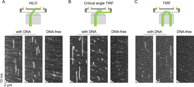
Figure 2.
Comparison of three illumination methods suitable for sub-millisecond resolved kymograph measurements of DNA-binding proteins. Typical kymographs of p53 in 150 mM KCl obtained at a time resolution of 500 μs based on the HILO (A), critical-angle TIRF (B), and TIRF (C) methods. The two kymographs on the left of each panel were obtained in the presence of the tethered DNA. The kymographs on the right were obtained without DNA. A schematic illustration of each of the illumination methods is shown in the top panels.

